


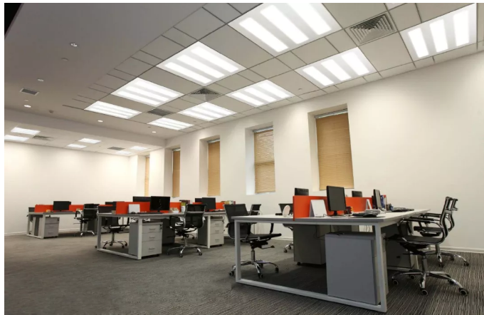
Industry Applications
This article summarizes the research and application of light-emitting diodes (LED) in animal models and clinical medicine in recent years, including the research of LED application in animal cells and human tissue cells, the research of LED application in animal models, and the experiment of LED in clinical treatment Research. Look forward to the prospect of LED application in clinical treatment. LED Phototherapy Cell Animal Clinical Light Emitting Diode (1ight emitting diode, LED) drive circuit is easy to control, has a long service life and is low in price. With the continuous development of semiconductor technology, LED has greatly improved the performance of parameters such as luminous intensity, peak wavelength, and half-wave bandwidth. At present, the light effect of a single LED has exceeded 1001mW; the peak wavelength is becoming more and more stable, the half-wave bandwidth is narrower, and the monochromatic performance is good; the directionality is good; the covering wavelength is from ultraviolet to infrared, and monochromatic LEDs of almost any wavelength can be found. These characteristics provide a technical basis for LEDs to be used in the medical field. This paper summarizes the work that LED has done in the medical field, and looks forward to the application prospect of LED in the biomedical field.
1 Research on the effect of LED on animal tissue cells Whelan et al. used LEDs of different wavelengths and irradiation doses to irradiate cells cultured in vitro. Cell proliferation increased by 55% to 71% compared with the control group. Whelan fibroblasts extracted from Sprague Dawley mouse skin were irradiated with 670nm LED at a dose of 4J/cm2, and osteoblasts (MC3T3-E1) were extracted from mouse bone with 670nm, 728nm and 880nm LEDs, respectively With a dose of 4J/cm2
On the second day after irradiation, the percentages of DNA synthesis in fibroblasts and osteoblasts were measured compared with those in the control group. It was found that the percentage of DNA synthesis in cells after LED irradiation was significantly increased, and at the second day The percentage of days is increased.
Liu Jiang et al. used different concentrations of simvastatin to culture mouse skeletal muscle C2C12 cells, and then irradiated them with red LEDs [wavelength (640±15) nm] of varying intensities for 2 days, 15 min/d. Cell proliferation was assessed by the methylthiazolyl tetrazolium colorimetric assay. It was found that the proliferation of C2C12 cells in the experimental group with a concentration of simvastatin of 2.0×10⁻⁵mol/L was inhibited, and this inhibitory effect could be antagonized by the photobiomodulation effect of red LED[1]. It is further confirmed that photobiomodulation only regulates dysfunctional cells or tissues, and has no effect on normal functioning tissues or cells.
For canine neuroma cells (2A9) and human glioblastoma cells (U373) cultured in vitro, use Lutetium Texaphyrin (Lutex, absorption peak 740nm ) and Benzoporphyrin Derivative (BPD, absorption peak 690nm ) as photosensitizers, using PDT method In the experiment of killing tumor cells, when LEDs with emission peaks at 728nm and 680nm were used as light sources, 50% to 86% of cancer cells in the two cell lines were killed.
2 Study on LED therapy in animals Whelan conducted a study on gene expression changes in traumatic hyperglycemic mice undergoing LED rehabilitation phototherapy. The drug was implanted subcutaneously in the back of the mouse, and LED phototherapy was performed every day. Finally, the skin was cut to remove the drug sponge to extract RNA, which was then analyzed using eDNA array. The results showed that compared with samples without phototherapy, gene regeneration was obvious in the samples after phototherapy. LED light therapy can accelerate the natural wound healing process, allowing patients to return to pre-illness levels of activity more quickly. In the experiment, Li Fengmin et al. used blue-violet light with a wavelength of 380-460nm, a peak value of 410nm, and an output power of 40mW to irradiate burn and trauma rat models. The irradiation distance was 8cm, and the spot diameter was 65mm.
3 Each irradiation dose was 0.72 J/cm2, 10 min, starting on the first day after operation, once a day, for 12 d continuously. Experiments have shown that blue-violet light in this wavelength range can significantly increase the content of hydroxyproline in rat burn wound skin tissue, thereby promoting the growth of granulation tissue and wound healing, and improving the tensile strength of the healing wound. Blue-violet light irradiation can increase the phagocytic rate and phagocytic index of peritoneal phagocytes in burnt rats, that is, improve the phagocytic ability of rat peritoneal phagocytes to improve the body’s immunity, thereby promoting wound healing [2].
3 LED research in clinical medical experiments
3.1 Treatment of acne Acne is a common sebaceous gland disease, with an incidence rate as high as 80% among adolescents. Excessive sebum secretion and proliferation of Propionibacterium acnes are the main causes of the disease. At present, local and systemic treatments are mainly based on antibiotics and isotretinoin drugs, but the use of antibiotics will lead to drug resistance of Propionibacterium acnes, and isotretinoin drugs will produce obvious side effects, resulting in limited clinical application. Propionibacterium acnes is irradiated with LED blue light with a wavelength of about 415 nm, which can cause the endogenous porphyrins (mainly protoporphyrin IV and coproporphyrin III) of anaerobic P. acnes to undergo a photochemical reaction to generate singlet oxygen. Thereby killing bacteria and achieving the effect of treating acne. LED blue light therapy is safe and effective without the drug resistance and side effects of traditional treatments.
3.2 Treatment of neonatal jaundice
The typical application of LED blue light phototherapy is the treatment of neonatal jaundice. The light that can be absorbed by bilirubin has the best wavelength of 450-460nm, while the main wavelength peak of LED blue light is between 425-475nm , so far it is still the best light source for phototherapy. In the experiment, Zhao Xinchun et al. randomly divided 120 neonates with clinical diagnosis of hyperbilirubinemia into
The experimental observation results of the LED phototherapy group and the luminal group: the clinical effective rate of the LED blue light phototherapy group was 83.3%, and that of the luminal group was 33.3%. Obviously, LED blue light irradiation is safer and more effective in treating neonatal jaundice.
3.3 Treatment of wound healing According to the research results of low-energy laser on wound healing, many companies have launched products that use LEDs to promote wound healing and tissue healing, and have done a lot of clinical research and obtained satisfactory results. Whelan and other studies on animal bodies and cells believe that LED irradiation can make human muscle and skin cells grow at 5 times the normal speed. The study was conducted by the Midwestern Athletes Against Childhood Cancer (MACC) Foundation Research Center at the Wisconsin-Madison School of Medicine. The FDA has approved the team led by WHELAN to conduct clinical trials on the experimental therapy of LED, and the results are satisfactory for wound recovery. At the same time, Whelan’s research is also used in tissue healing in a gravity-free environment, applying this technology to long-distance and long-duration space travel to treat astronaut injuries. In addition, the technology was used on a US submarine with an array of LEDs to heal potential training injuries. 3.4 Increasing the body’s endurance and anti-fatigue ability Jia Danbing et al. used LED red light with a wavelength of 600nm-1200nm to detect the content of myocardial enzymes, blood coagulation series and blood routine related molecules, and studied the effect of red light irradiation on human body’s anti-fatigue ability and Endurance effects. Results Compared with the control group, the contents of HB, IBC and Ca in the experimental group were significantly increased after red light irradiation, while other indicators did not change significantly, and HB and Rt3C played an important role in tissue oxygen supply. During the body’s exercise, by improving the tissue’s ability to withstand hypoxia, reducing the anaerobic metabolism of the tissue and reducing the generation of lactic acid during exercise, the body’s soreness and fatigue can be significantly reduced under the same intensity of exercise, and the body’s endurance and resistance can be improved. Fatigue ability. The experiment showed that the content of HB and 5 after red light irradiation increased significantly compared with that before irradiation through self-control and inter-group control research. After light irradiation, the oxygen-carrying capacity of the blood is significantly increased, and the body’s endurance and fatigue resistance are increased.
3.5 Oral Ulcer Inflammation Treatment
Some pediatric patients with leukemia receive maximal doses of chemoradiotherapy to kill the neoplastic bone marrow before implanting bone marrow that matches their cellular antigens, as chemotherapy drugs and radiation therapy would not Differentially kills rapidly dividing cells, such as oral mucosal cells and gastrointestinal cell lines. These chemotherapy patients often cause acute oral ulcers, resulting in difficulty in swallowing and swallowing, and severe gastrointestinal reactions. Using LEDs with a wavelength of 688nm , after the last chemotherapy, they received a LED irradiation dose of 4J/cm2. Among the 30 patients treated with LEDs, 47% of the oral pain was relieved, and the degree of healing of their oral ulcers was better than expected better.
3.6 Treatment of seasonal depression
One of the best ways to beat the seasonal blues is to use light. Light can stimulate the retina, and the retina transmits this stimulus information to the nervous system, and then to the light-sensitive melatonin in the brain. Melatonin plays an extremely important role in adjusting the body’s circadian rhythm. Patients with seasonal depression have elevated levels of melatonin. Because full-spectrum light reduces melatonin secretion, it is very effective in treating SAD patients with light. Green light (wavelength 540nm) is the most obvious spectrum to inhibit melatonin secretion. In 1991, IBERMAN mentioned that LEWY believed that irradiating with white light with an intensity of 10mW for 30 minutes after getting up every day can inhibit the secretion of melatonin and relieve seasonal depression. This observation is important not only for understanding the physiological consequences of melatonin, but also for regulating secretion rhythms.
6 SAD and other health problems. At present, relevant patents on the application of LEDs have been applied for, and the content is light therapy for various physiological and mental disorders, such as light therapy for SAD.
【References】Photobiomodulation of C2C12 Cell Proliferation by Red Light Emitting Diode Irradiation[J]. Chinese Clinical Rehabilitation, 2006, l0(13): 107-109
Effects of blue-violet light on skin wound and burn healing in rats. Chinese Journal of Physiotherapy [J]. 2001, 24 (5): 294-295.
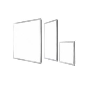
Recommended specifications
optical instrument
optical instrument
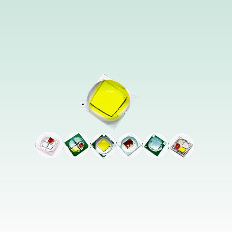
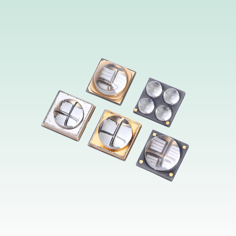
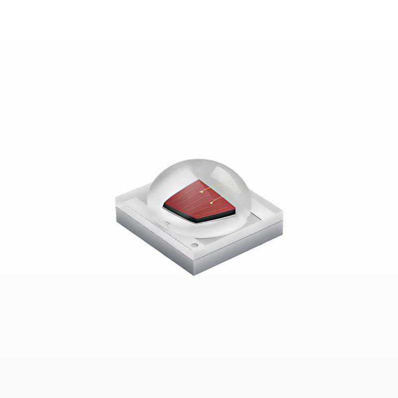
High-Power LED
High-Power LED
Explore More from Queendom Lamp
Stay updated with the latest LED technology, lighting solutions, and industry insights.
Request a Quote About Queendom